Nanoscopic Characterization of Cellular Processes
This TIMed CENTER Core Facility investigates the biomolecular mechanisms underlying biology and medicine.

In order to realise this project, high-resolution single-molecule fluorescence microscopy (SM-FM) is sometimes used. This is utilised to observe specific molecules in living cells, tissues and entire organisms. Fluorescence microscopy can be used to investigate biomolecular and cellular dynamics, co-localisations and interactions. The basis for this is the selective and specific labelling of certain cell components.
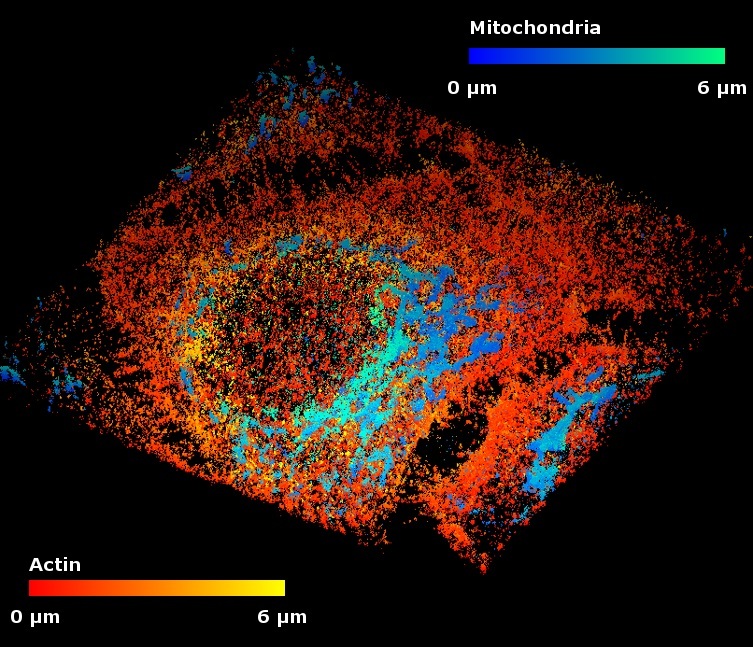
Another important instrument of this Core Facility is super-resolution microscopy (PALM/STORM). This is a technique used to represent fixed or living cells and different kinds of tissue in a three-dimensional space. The series of images generated in this way are transferred to a film sequence for analysis. The multiscale parameters of dynamic and static cellular processes are then analysed using special software packages. In this way, processes can be recorded and quantified in a computerised and automated manner. In addition to the imaging process, molecular biological techniques are used to characterise biomolecules.

Functions
- Real time-visualization of biomolecules, interactions and dynamics
- (Real time) analysis of dynamic and static cellular and biomolecular processes (diffusion, localization, morphology, protein cluster) by means of specialized software packages
- 3D-localization of biomolecules in cells and tissue by means of super resolution fluorescence microscopy
Services
- Determining affinities, stoichiometry, multivalence, interaction kinetics of molecules, absorption of molecules by cells
- Proof of proteins/RNA/DNA in cells from cell cultures in tissue
- Tests of bio-markers (e.g. fluorescence markers)
- Toxicity tests (e.g. on surfaces
Contact
I'll help you choose your study program.