Equipment and Software
In 2004, the first 3DCT device in Austria was installed at the Upper Austria University of Applied Sciences - Campus Wels. Meanwhile, the CT research group has four CT-systems.
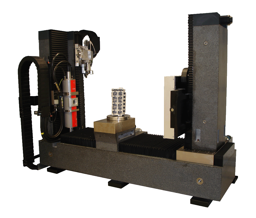
Procon CT-Alpha
The Dual Source computed tomograph CT-Alpha was installed at the FHOÖ Wels Campus in November 2004 and upgraded to the current state in 2022. The device has two X-ray sources: the microfocus X-ray source X-Ray WorX XWT-240-CT Plus is suitable for high-resolution measurements, the minifocus X-ray Comet MXR-451HP/11 for high penetration lengths. The detector implemented is an a-Si (amorphous silicon) detector type PerkinElmer XRD 1620 AN14 CTS converting X-rays via a scintillation layer into visible light. The technical data on the device is listed below:
- 240-kV-microfocus- and 450-kV-minifocus tube
- 2048 * 2048 pixel 16 bit a-Si flat panel detector
- Minimum voxel size: 5 µm (max. sample diameter / 2048)
- Sample weight < 80 kg
- Sample diameter < 310 mm (with scan range enlargement < 600 mm)
- Sample length <1,8 m
- Maximum penetration length microfocus 240 kV: polymers - max. 200 mm, aluminium - max. 120 mm, steel - max. 30 mm
- Maximum penetration length minifocus 450 kV: polymers - max. 500 mm, aluminium - max. 250 mm, steel - max. 70 mm
- "Region of Interest" measurement mode for flat components
- Radioscopy mode with up to 7 fps
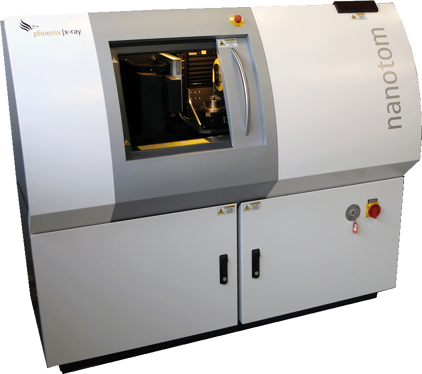
Phoenix/X-Ray Nanotom 180
The high-resolution Sub-µm computed tomography device phoenix|x-ray (Part of GE Sensing and Inspection Technologies) Nanotom 180 has been started up in February 2008. The system includes a nanofocus X-ray source with transmission target and a max. acceleration voltage of 180 kV. The Nanotom detector type Hamamatsu C7942SK-05 allows for a scanning space with a resolution of 2300 * 2300 pixels.
Detailed technical data of the Sub-µm device is listed below:
- 180-kV-nanofocus tube with minimum focal spot diameter of < 1 µm
- 2300 * 2300 pixel 12 bit Hamamatsu detector
- Triple-scan range enlargement mode
- Minimum voxel size: 0,5 µm (max. sample diameter / 2300)
- Sample weight < 2 kg
- Sample diameter < 68 mm (with scan range enlargement < 210 mm)
- Sample length< 150 mm
- Maximum penetration length: polymers - max. 50 mm, aluminium - max. 30 mm, steel - max. 4 mm
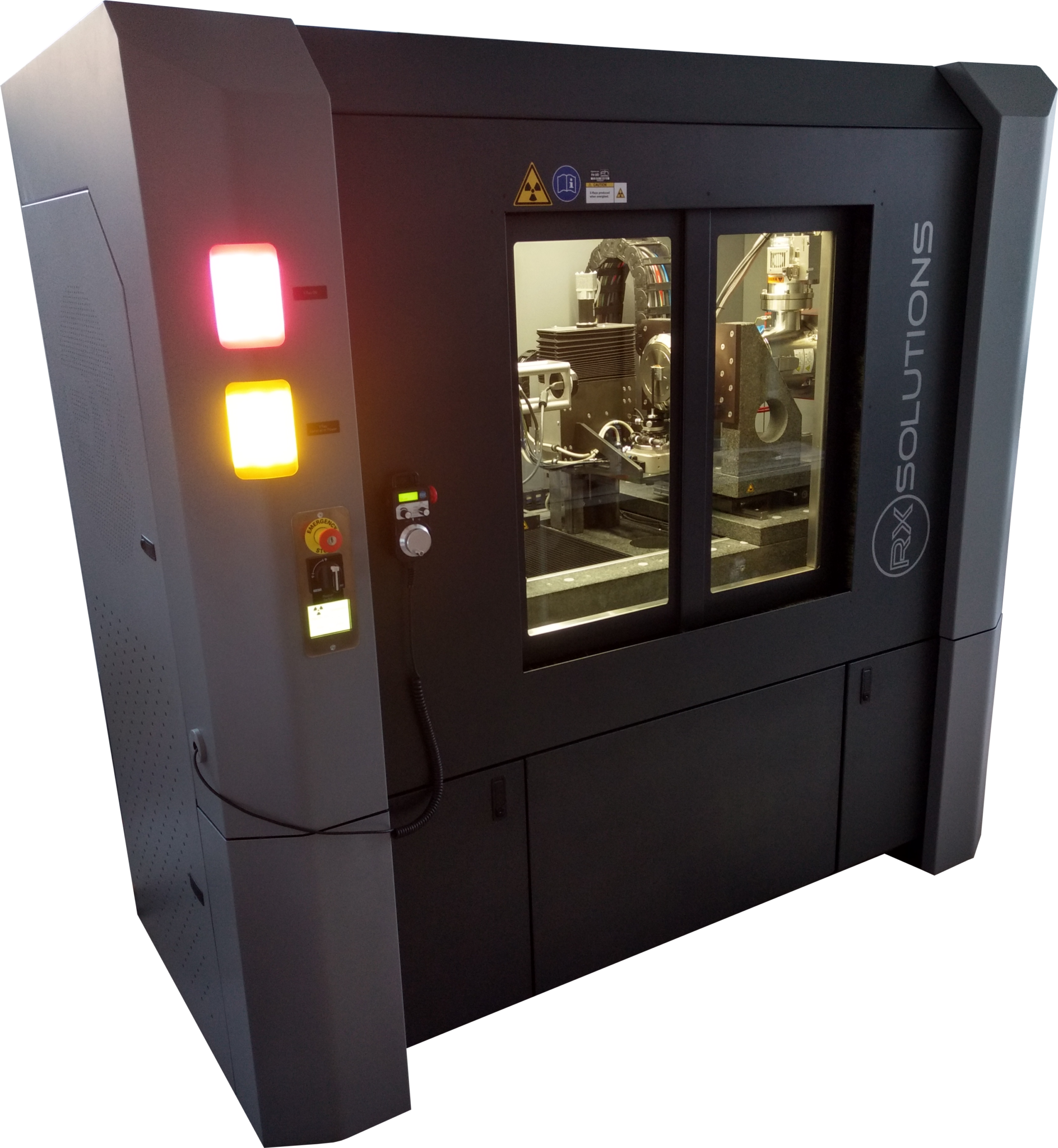
RX Solutions Easytom 160
The high-resolution nano computed tomography device EasyTom 160 from RX Solutions has been started up in April 2017. The system includes a nanofocus X-ray source with transmission target and a max. acceleration voltage of 160 kV, which can be mounted with tungsten or LaB6 filaments. The nano device can be operated either with a flat panel detector with 1920 * 1536 pixels for higher energies and faster data acquisition or with a CCD camera with 4008 * 2672 pixels for lower energies and higher resolution.
Detailed technical data of the device is listed below:
- 160 kV Hamamatsu nanofocus tube with focal spot diameter of < 400 nm (LaB6 filament)
- 1920 * 1536 pixel 16 bit Varian flat panel detector
- 4032 * 2688 pixel 14 bit CCD camera
- Measurement of materials with low density < 1 g/cm^3
- Triple-scan range enlargement mode
- Minimum voxel size: 50 nm
- Sample weight < 20 kg
- Sample diameter < 200 mm
- Sample length < 700 mm
- Maximum penetration length: polymers - max. 50 mm, aluminium - max. 30 mm, steel - max. 4 mm
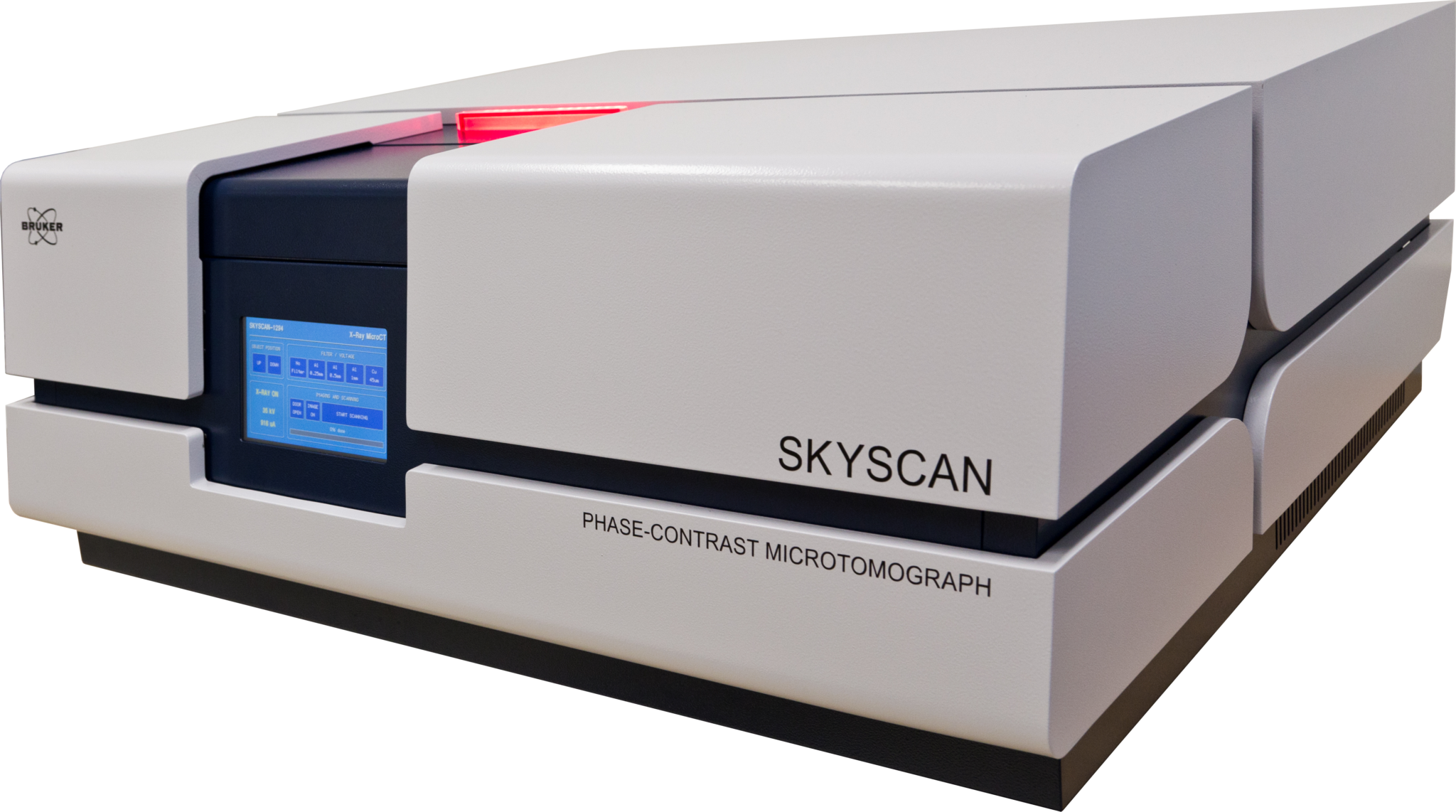
Skyscan 1294
The SkyScan 1294 desktop phase-contrast X-ray microtomograph was installed at the FH OÖ Campus Wels in January 2015. The system includes a specially designed micro-focus X-ray source, a 11 megapixel X-ray camera and a precise three-grating Talbot-Lau X-ray interferometer for simultaneous extraction of absorption contrast, differential phase contrast and dark-field images. This phase-contrast system will primarily be used for detailed investigation of polymeric and biologic material systems. The technical data on the device is listed below:
- 60-kV-microfocus tube with a focal spot diameter of 33 µm
- 4000 * 2672 pixel 12 bit detector
- Measurement of materials with low density < 2 g/cm³
- Minimum voxel size: 5.7 µm
- Sample diameter < 20 mm
- Sample length < 60 mm
Software
The following means of evaluation are at the research groups disposal:
- VG Studio Max with additional modules
- MAVI
- IAR (Iterative Artefact Reductions) software
- Actual-nominal comparison tools
- Zeiss Calypso with CT-extensions
- Our in-house developed tools
FHOÖ Equipment
Aside the afore mentioned computed tomography devices, the extensive and modern university equipment includes the following technologies:
- Serial sectioning Tescan Vega / Oxford
- Scanning electron microscope with X-ray microprobe for chemical analysis
- Testing equipment for active thermography
- Testing equipment for shearography
- Optical scanner GOM Athos
- Coordinate measurement device Zeiss Spektrum 7/7/6
- Rapid prototyping device:
- 3D-printer (Z-Cooperation)
- Laser melting / -sintering device (Concept)